Practical 3: Symptom, Isolation and Pathogenecity Test
Activity 1: Identification of plant disease through signs and symptoms observation
OBJECTIVES
1. To identify plant disease through signs and symptoms
2. To learn how to describe signs and symptoms of plant disease.
METHODS
1. The sample of symptoms of plant disease is observed.
2. The symptoms are described and host plant and causal agent are identified.
3. Slides to observed causal agent under light microscope are prepared.
RESULTS
1) Plant disease symptoms
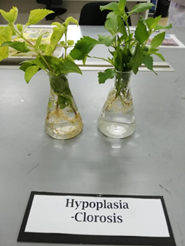
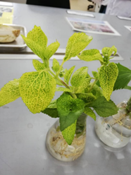
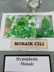
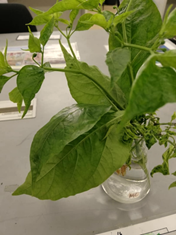
Hypoplasia


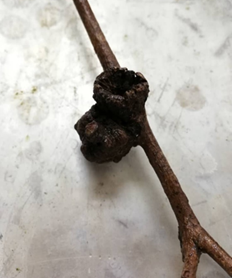
Hyperplasia

Hypertrophy

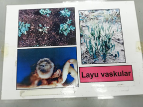
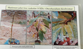
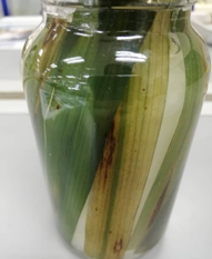
General Necrosis
Local Necrosis


DISCUSSION
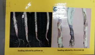
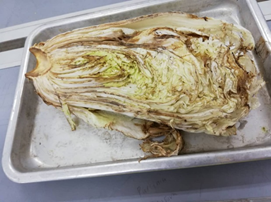
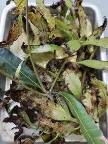
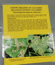
There are many types of disease symptoms that occur in plants. The symptoms include general necrosis, local necrosis, hypoplasia, hypertrophy and hyperplasia. Necrosis often describes as death of cells or tissues and can be divided into general necrosis and local necrosis. General necrosis is more general and affect all part of the host. Examples of general necrosis are damping-off, blight, blast, dieback, vascular wilt and soft rot. Besides, local necrosis is more limited to only cells surrounding the invading pathogen. The examples are anthracnose, powdery mildew, downy mildew, canker, and rust.
Furthermore, disease symptom hypoplasia involves the reducing size of cells and plant growth retarded. The plant experience under development which include stunting of leaves, shortened internodes and inadequate chlorophyll production. It is caused by many types of pathogen and the examples include chlorosis and mosaic. Moreover, hypertrophy and hyperplasia are symptoms for overgrowth. Hypertrophy involves cell enlargement that leads to symptoms such as smut and gall. Hyperplasia involves fast cell division that results in symptom called witches broom.
Activity 2: Koch’s Postulates
OBJECTIVES
- To observe disease symptoms of the casual agents on chilli parts.
- To determine pathogenicity of the causal agent of the plant disease on chilli, whether Collectotrichum truncatum or Collectotrichum capsici by Koch’s postulate.
- To observe the presence of disease on chilli
MATERIALS
Pure culture of 2 fungal pathogen specimen (Collectotrichum truncatum and Collectotrichum capsica), diseased and healthy chilli fruit, petri dish, filter paper, PDA plates, forceps, inoculation needle, plastic container, slides, spirit lamp, scalpel, coverslips, microscope and Lactophenol Cotton Blue (LCB)
METHODS
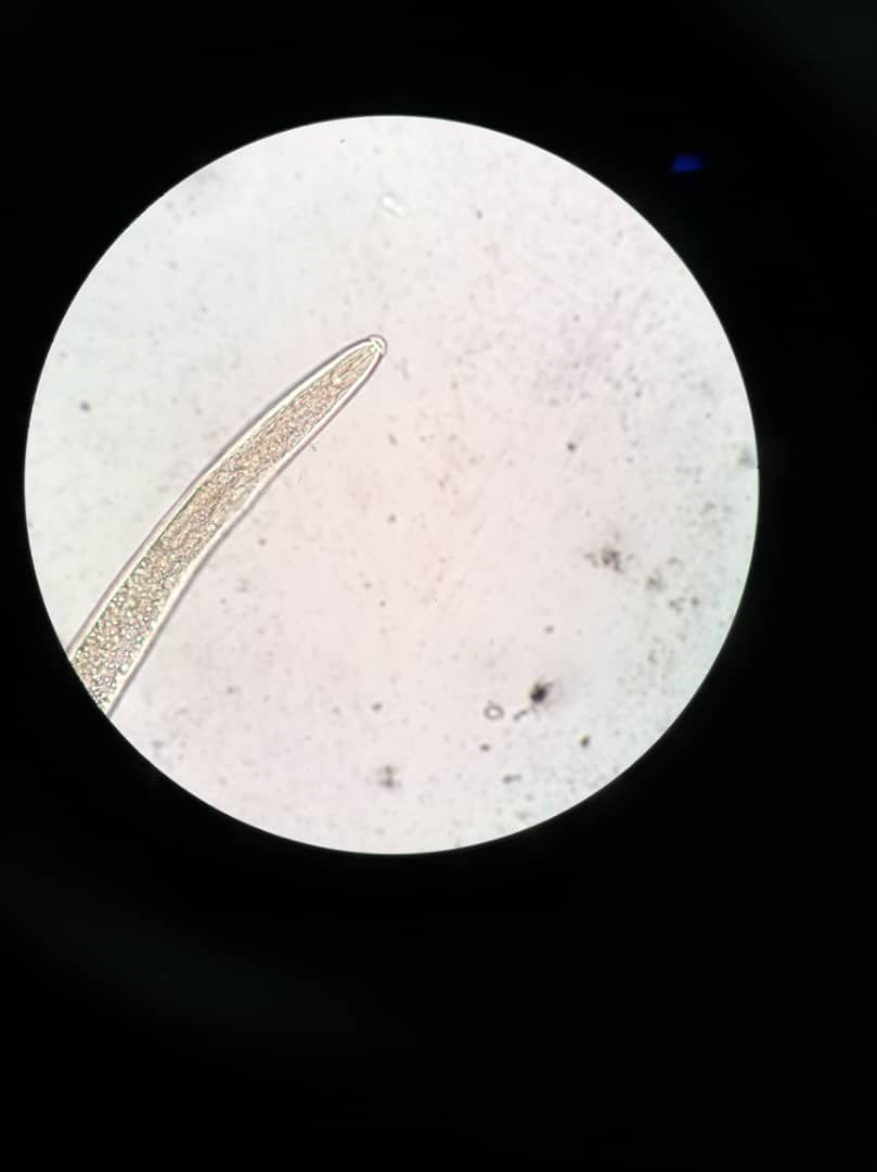
- Slides from both cultures are prepared and observed under light microscope.
- The causal agent is isolated from the disease tissues onto PDA (Potato Dextrose Agar) plates. The plates are labelled and incubated at room temperature for observation.
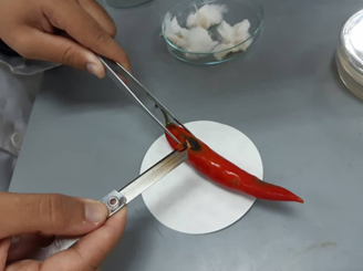

- Aseptically, 1 agar containing hyphae is cut from each pure culture provided and being inoculate each species of pathogens on sterilized chilli fruit. Third chilli is uninoculated and used as control. The fruit is kept in moist tray and is covered with plastic sheet. The fruit is incubated for 3-5 days and the symptoms exhibit is observed.


- The isolation process as in step 2 above is repeated using fruit that demonstrate similar symptoms as in step 2 above.
RESULTS
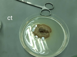
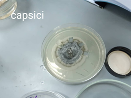
- Inoculation from disease infected chili into PDA plates
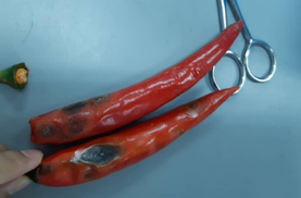
Symptoms on specimen provided shows black swirling, dieback ripe fruit rot and brown necrotic lesions.


The result shown characteristic of Collectotrichum capsici
- Koch’s Postulates
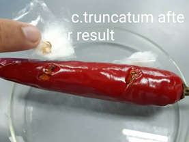
Chili infected by Collectotrichum truncatum
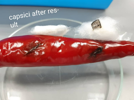
Chili infected by Collectrotrichum capsici

Controlled chili
DISCUSSION
In the experiment, we use two cultured fungi which
is Collectotrichum truncatum and Collectrotichum
capsica to carry out Koch’s Postulates. These two fungi are in the same
genus Collectotrichum sp. Koch’s Postulates that we carry out involve
inoculation and isolation. Isolation method is method to isolate suspected
causal agent from disease host plant and grown in pure culture. Inoculation
method involve pure culture of the suspected causal agent is inoculated into
healthy susceptible host plant. The host must able to shows the symptoms of the
suspected disease.
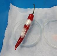
In the result, it shows that after 5 days, healthy
chili that infected by Collectrotichum capsica shows
the same symptoms as the infected chili specimen provided which is black
swirling spots, dieback ripe fruit rot and the development of brown necrotic
lesions around the black spot began to take place. The infection shows invasive
and severe. While healthy chili that is infected by Collectotrichum
truncatum shows only mild infections on the injured area. Therefore,
chili fruit is more susceptible host to causal agent Collectrotichum capsica
than Collectotrichum truncatum.
CONCLUSION
In conclusion, the disease symptoms of the causal
agent shown on the chili are black swirling spot, dieback ripe fruit rot and the
development of brown necrotic lesions around the black spot on the fruit.
Moreover, from the Koch’s Postulates procedure, we could determine the
pathogenicity of fungus Collectrotrichum capsici that infect the healthy
chili. The disease that cause the symptoms shown on the infected chili is
anthracnose disease.

Comments
Post a Comment