Practical 7: Phytobacteriology
Activity 1: Morphology of Bacteria
OBJECTIVES
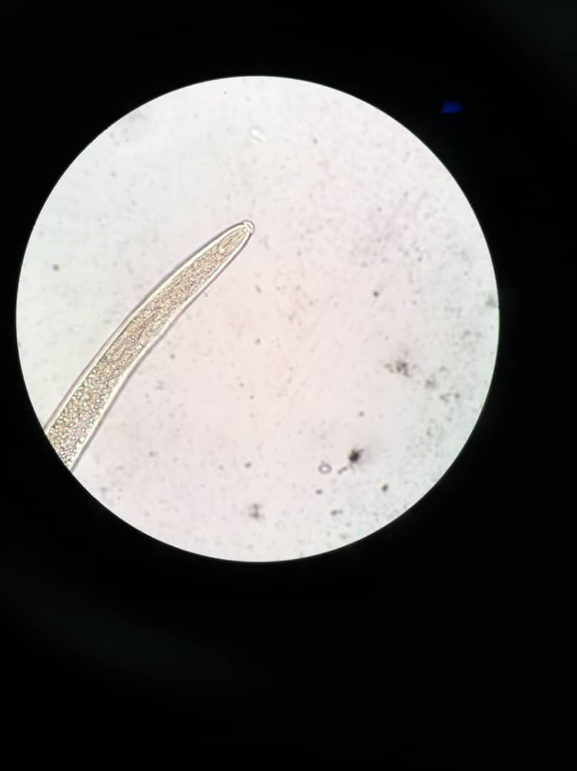
- To observe the morphology of bacteria on slides provided under microscope.
- To observe the shapes of bacteria colonies produce on NA plates.
- Different shapes of bacteria


- Bacteria colonies on NA plate

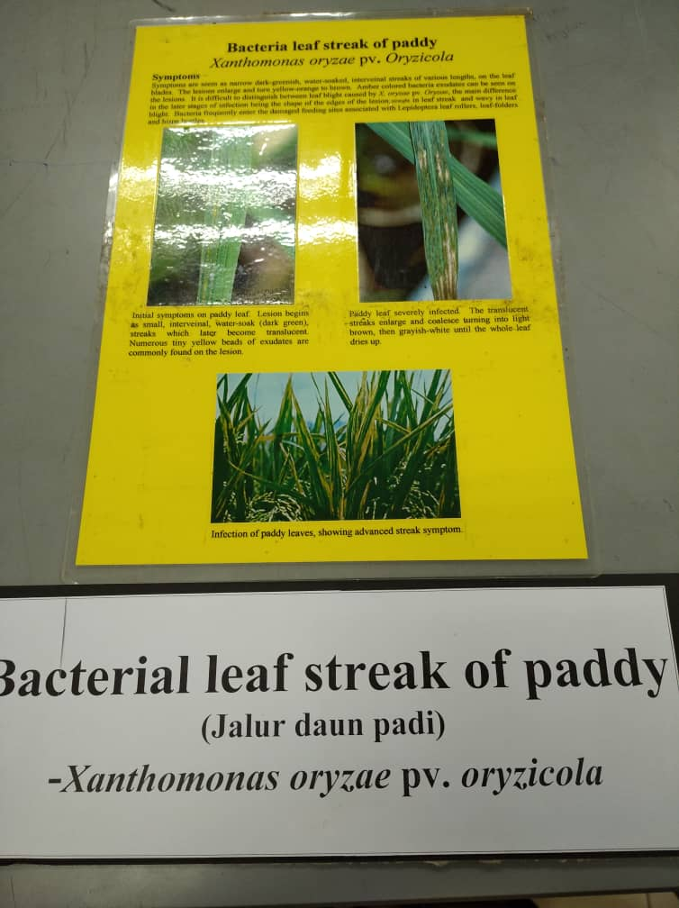
Activity 2: Disease Symptoms
OBJECTIVE
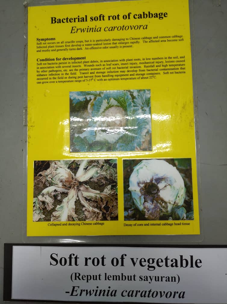
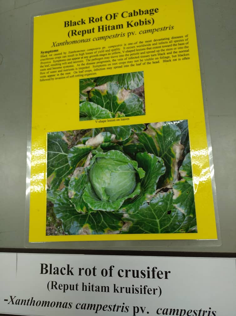
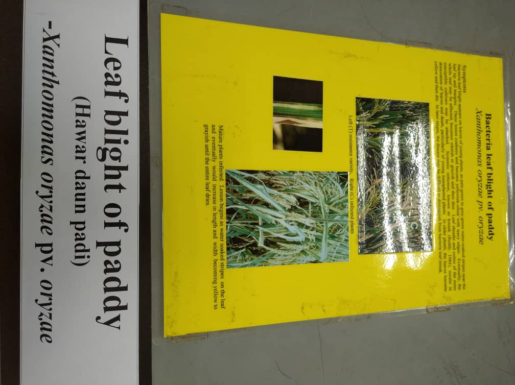
- To observe the symptoms caused by bacteria on the speciimen provided
Disease Symptoms


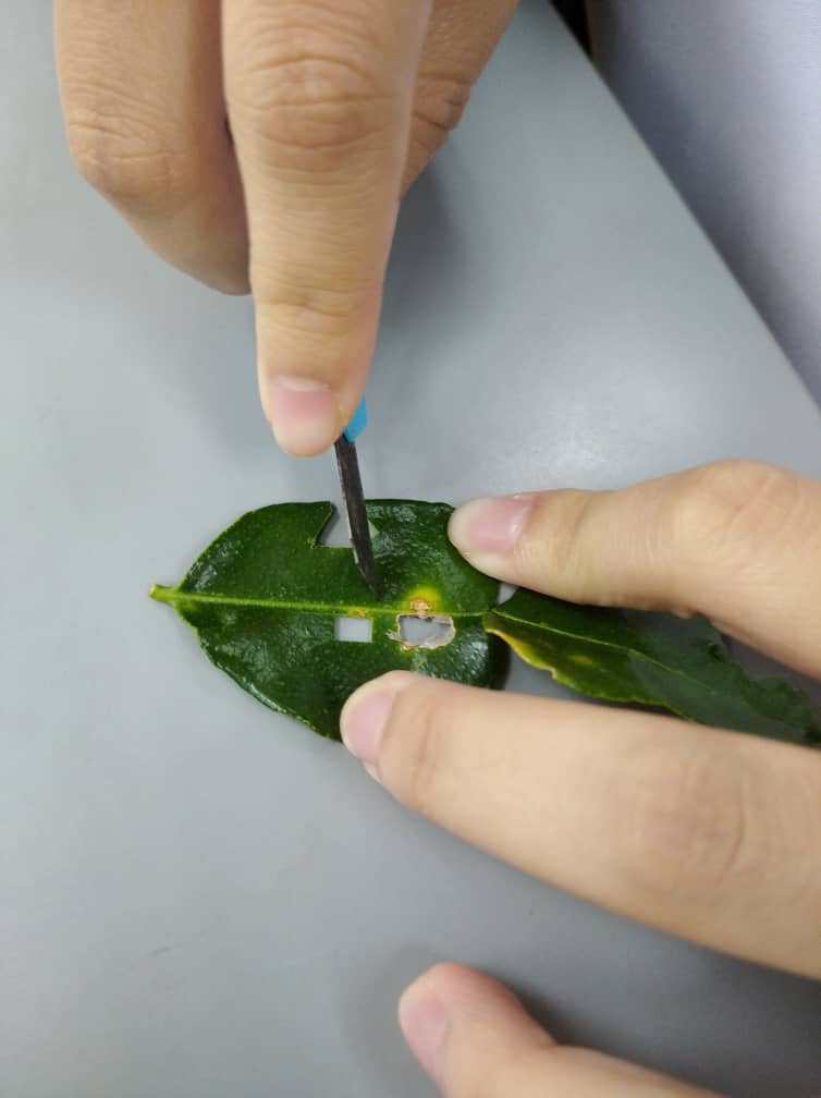
Activity 3: Bacteria Isolation from Plant Tissue
OBJECTIVE
- To learn techniques on how to isolate bacteria from plant tissue.
MATERIALS
Spirit lamp, petri dishes, inoculation needles, filter paper, pen knife, 70% alcohol, 10-20% chlorox, distilled water, citrus leaves and potato.

METHODS
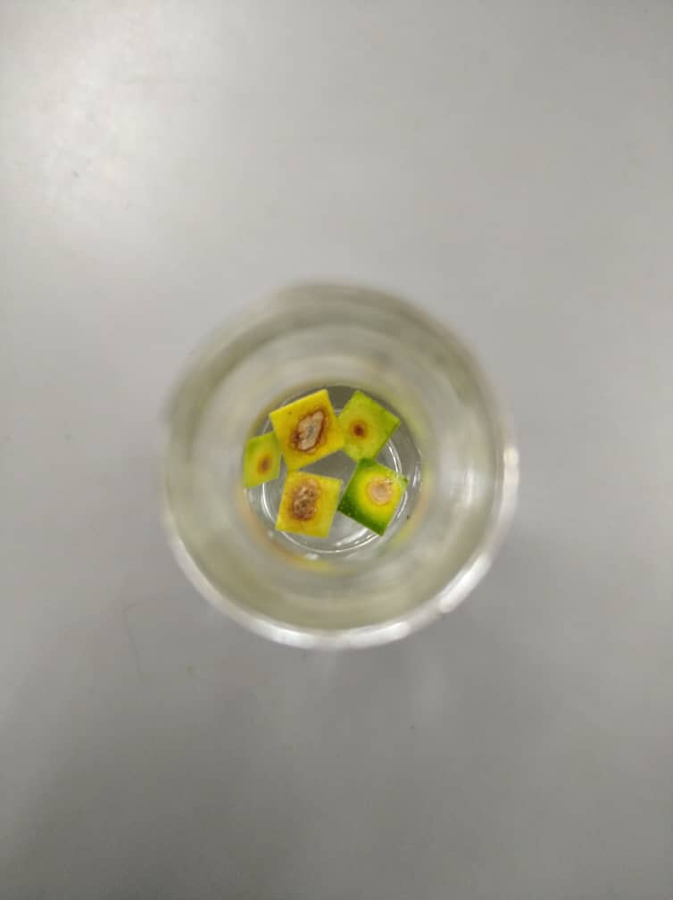
- Direct Isolation
- Pen knife is wipe with 70% alcohol and 10-20% chlorox and used to cut canker spots from disease tissues.
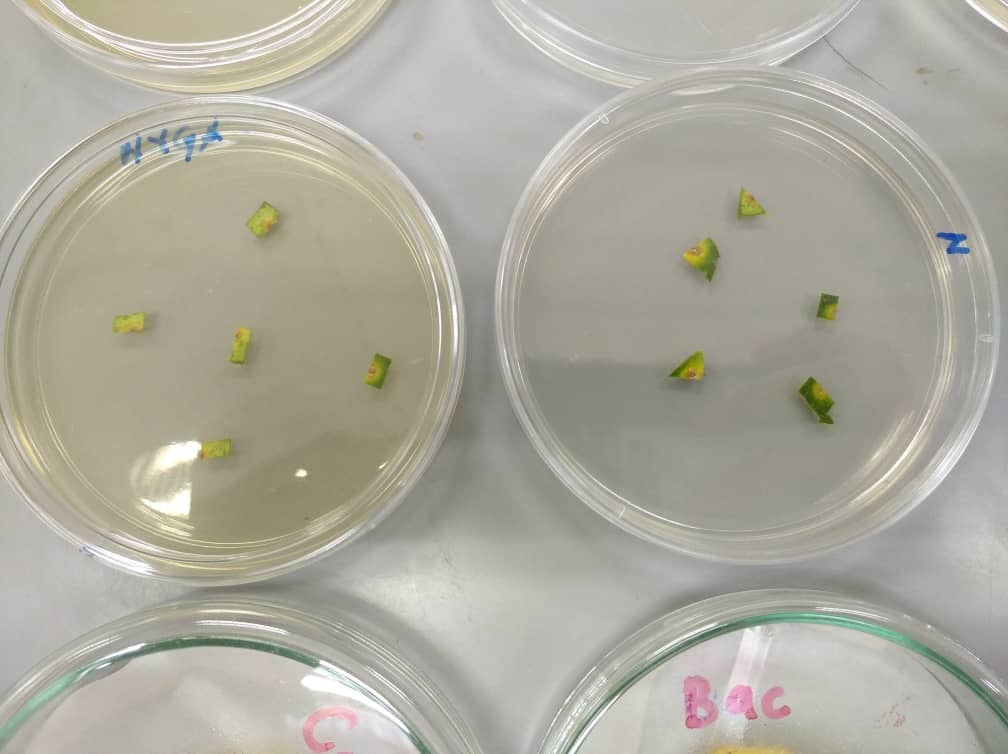
- The cut tissues are sterilized and placed in NYGA or NA media. The plates are incubated at 27-30 degrees celcius for 24-48 hours.

b) Dilution Streak
- Some of the cut out diseased tissues from earlier experiments in (a) is crushed in distilled water.
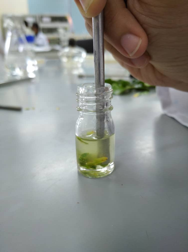
- Dilution streak is done on the provided petri dish plates containing appropriate medium.

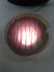
RESULTS
Dilution streak result and direct isolation.


The result shows that bacteria Xanthomonas axonopodis that were diluted from the plant tissue reproduce rapidly in the area that the streak is done.
Activity 3: Diagnostic Test Using the Cut Plant Tissues
OBJECTIVE
- To learn technique to isolate pathogen on plant tissue to confirm diagnosis.
- To observe the ability of bacteria to soften the plant tissue and cause rot.
MATERIALS
Spirit lamp, petri dishes, inoculation needles, pen knife, 70% alcohol, 10-20% chlorox, distilled water, forceps and potato.
METHOD
- Potato is washed, the skin is peeled off.
- Diseased slices 7-9mm thick is cut out and arranged in sterilized Petri dish.
- The “V” shaped is cut on the slice and a drop of sterile water is put on the slice. This slice is labelled as control.
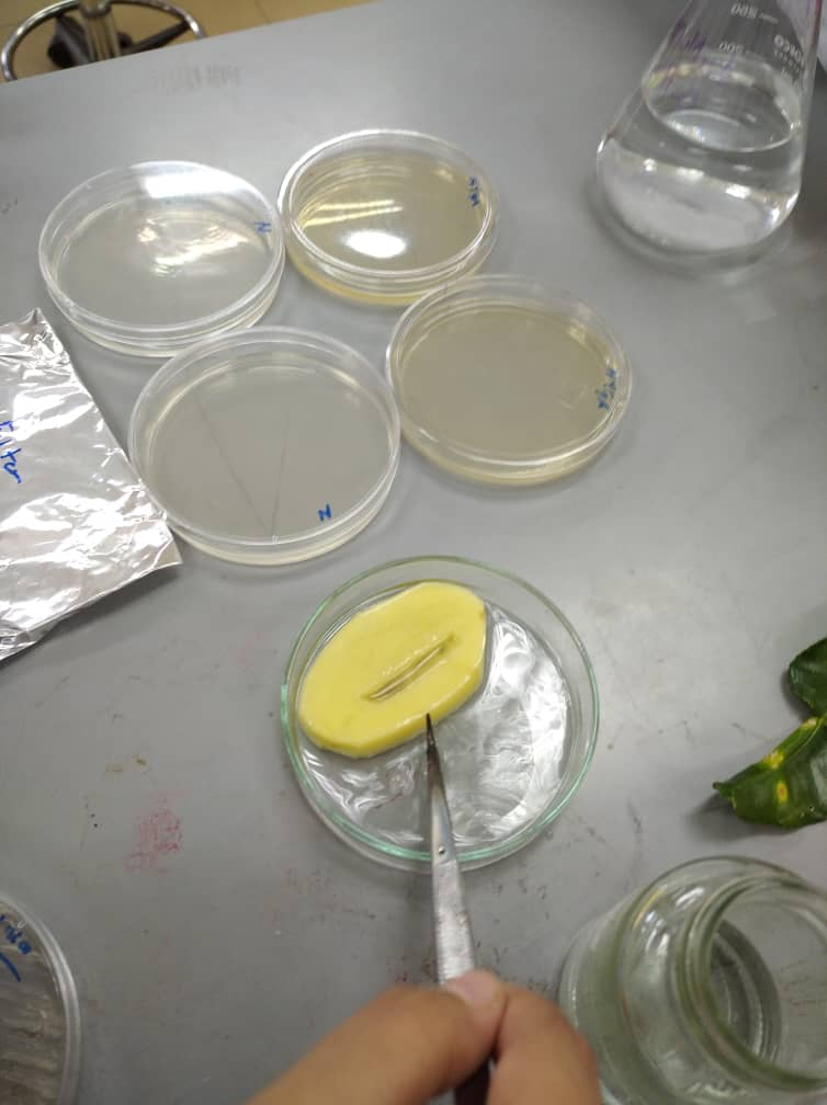

- The “V” shaped is cut on another slice and Erwinia caratovora subspecies caratovora is inoculate on the slice.

- All the plates in incubated for 24-48 hours AT 30°C. Symptoms is observed.
RESULT
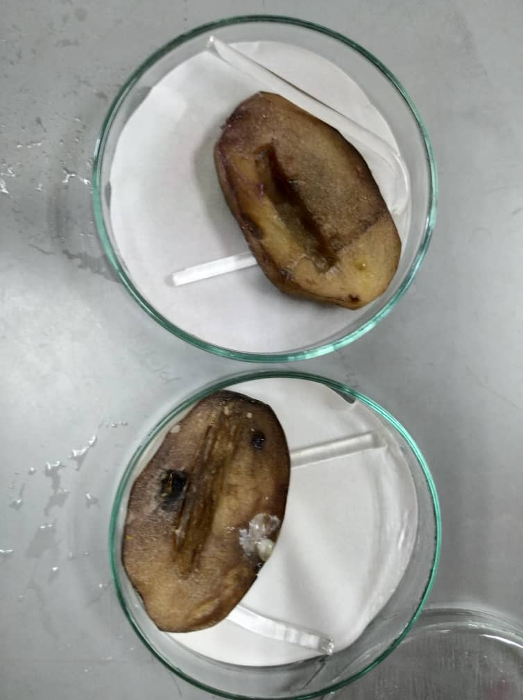
On the left side is the result for potato slice that act as control and on the right side is the result for the inoculation of Erwinia caratovora subspecies caratovora
DISCUSSION
The result shows that potato sliced that are infected by Erwina caratovora shows symptoms of soft rot. On the surface of potato seems like wet and mushy due to the infected tissues that become soft. The colour of the infected tissues turns dark brown or black. The bacteria first infect around the cut area and then spread around it.
CONCLUSION
In conclusion, the symptoms of soft rot bacteria is the infected tissues turned watery, rot and produce bad odor. It will eventually turn from healthy normal colour into dark brown or black. It is proven by inoculating Erwina caratovora on the potato sliced that shows the same symptoms as stated. Moreover, we also concluded that this diagnostic test using cut plant tissues is an easy method to be done and observe the outcomes accurately.

Comments
Post a Comment